
Guide · 12 min read
Mobile X Ray and Ultrasound at Home
The radiator clicks as an adult daughter sits beside her father's bed, phone pressed to her ear. He's 82, he fell getting out of the shower three days ago, and his hip still hurts. The family keeps discussing the same problem: arranging wheelchair transport, waiting in a hospital radiology lobby, and getting him safely home may feel harder than tolerating the pain. So far, he hasn't gone.
This is the gap mobile X ray and ultrasound can help address. Portable imaging brings the equipment to a patient who may not be able to reach a hospital easily, while a clinician connects the scan to the person's symptoms, medications, and goals of care. For families searching for Visiting physicians NJ, Visiting Doctors NJ, or Doctor home visits for elderly, the important distinction is that imaging works best as part of coordinated in-home medical care, not as an isolated test ordered without context.
Table of Contents
- Bringing Hospital Imaging to the Living Room
- What Mobile X Ray and Ultrasound Actually Are
- How the Two Modalities Compare at Home
- How an At-Home Imaging Visit Works Step by Step
- Who This Helps and Where It Falls Short
- How Life Primary Care Coordinates Mobile Imaging
- Practical Questions Families Ask Before Booking
Bringing Hospital Imaging to the Living Room
A technologist arrives with equipment that is genuinely portable. A mobile X-ray unit can be positioned near a bed or chair, while an ultrasound professional can examine the patient at the bedside. The images are then sent for interpretation by a radiologist, so the visit doesn't end with a guess made in the living room.
For the father after the fall, a house call clinician might first ask how he landed, examine his hip and leg, review his medications, and assess whether he can safely stand. If an X-ray is appropriate, the clinician can arrange imaging without making the family transfer him into a car or ambulance just to obtain a first diagnostic answer. A suspected major injury or emergency still requires hospital care, but many non-emergency questions can begin at home.
A 2025 systematic review of home-based diagnostic imaging identified 26 studies published between 1998 and 2023 across 15 countries. It found that X-ray imaging and ultrasonography were the main at-home diagnostic tests, most often used for patients with mobility limitations, supporting the view that home imaging is an established part of care rather than a novelty. The review is discussed in the context of services provided by mobile imaging specialists.
Practical rule: The value of home imaging isn't only convenience. It's the chance to obtain a useful answer without allowing transportation barriers to decide whether a patient gets assessed.
Mobile X-ray has particular value in nursing homes, homes for older adults, hospice settings, shelters, and other non-hospital locations where transport creates delays. One review describes good image quality and potential cost effectiveness outside the hospital, while noting that taking the source and detector to the patient can remove ambulance or wheelchair transport delays and shorten time to diagnosis in suitable cases. Families arranging House calls for seniors can learn more about how care is delivered through this overview of healthcare delivered at home.
If the patient does need a hospital transfer, reducing confusion at registration can also help. A practical guide to smoother hospital front desk explains ways families can prepare information and communicate more clearly during a stressful arrival.
What Mobile X Ray and Ultrasound Actually Are
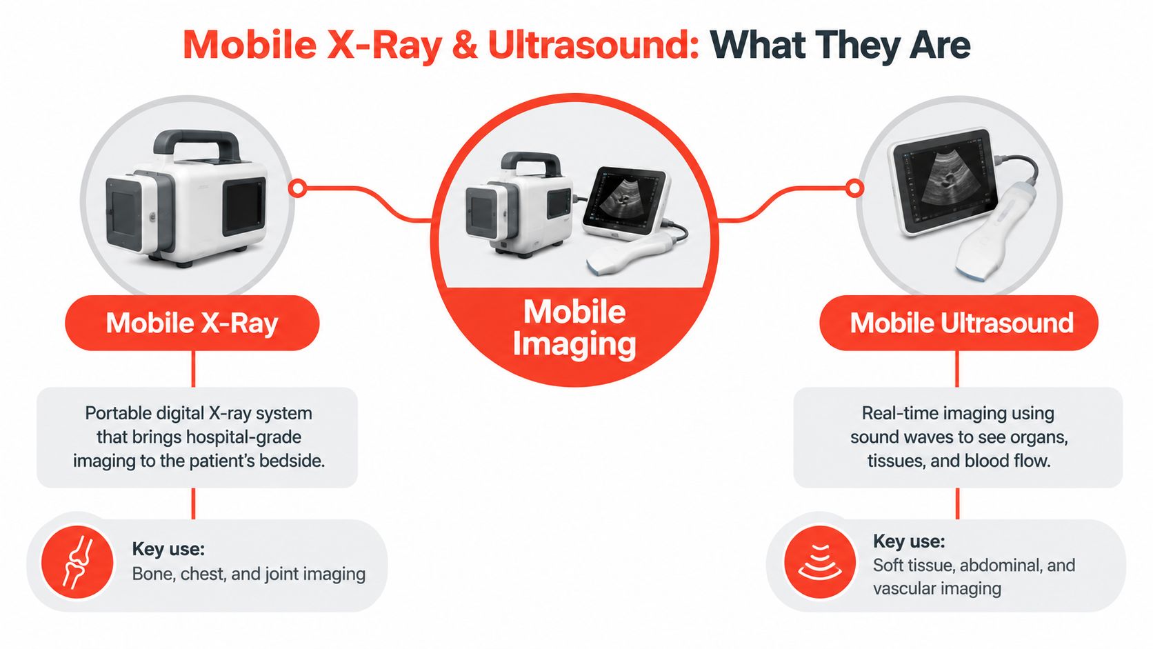
Start with the simplest distinction. Mobile X-ray uses X-rays and a digital detector to create still images, especially of dense structures such as bones and the chest. Ultrasound uses high-frequency sound waves to create images in real time, making it useful for many soft tissues, organs, fluid collections, and blood-flow questions.
Mobile X-ray in plain language
Think of an X-ray as a pattern made by invisible electromagnetic waves passing through the body. Bone absorbs more of the energy than softer tissue, so the detector records a contrast pattern that can show fractures, lung changes, and other abnormalities. A portable digital unit performs the same basic type of radiography used in a hospital, but the technologist brings the source and detector to the patient.
Portable home imaging can cover areas including the chest, spine, pelvis, knees, shoulders, arms, hands, legs, and feet, according to a description of home imaging services from portable X-ray and ultrasound diagnostics. The exact body part depends on the order and the equipment available.
Ultrasound as medical sonar
Ultrasound works more like submarine sonar. The technologist moves a probe over the skin, and sound waves travel into the body, bounce back from different tissues, and return as echoes. The system converts those echoes into moving images, sometimes including views of blood flow.
Ultrasound doesn't use ionizing radiation, and neither modality generally requires sedation or a contrast injection for the basic studies described here. A credentialed technologist performs the examination, then sends the images to a radiologist. The technologist may confirm that the images were captured adequately, but the formal interpretation comes from the radiologist and is returned to the ordering clinician.
Readers who want a broader look at sound-based imaging can explore acoustic imaging tagged articles, while keeping in mind that an educational article can't determine which scan a particular patient needs.
How the Two Modalities Compare at Home
Mobile X-ray and mobile ultrasound aren't competing versions of the same test. They answer different clinical questions.
A fall with concern for a broken hip, rib, or spine often points toward X-ray because bone absorbs X-rays clearly. A swollen leg, suspected fluid around an organ, or a question involving soft tissue may call for ultrasound. A chest X-ray can help evaluate certain lung or heart-related findings, while ultrasound can provide real-time views of selected organs, tissues, and blood flow.
Portable ultrasound has become easier to use outside fixed radiology departments because modern devices are lightweight, battery-powered, and transportable. In one comparative study summarized by research on imaging access in remote settings, handheld ultrasound showed 92.9% sensitivity, 92.3% specificity, and 92.5% accuracy, compared with 77.8% sensitivity, 91.5% specificity, and 89.3% accuracy for a cart-based system in that comparison. Those figures apply to the reported study, not to every patient, device, or operator.
| Feature | Mobile X Ray | Mobile Ultrasound |
|---|---|---|
| Main signal | X-rays captured by a digital detector | Sound-wave echoes captured by a probe |
| Strongest general role | Bone, chest, and selected abdominal imaging | Soft tissue, organs, fluid, and blood-flow imaging |
| Radiation | Uses ionizing radiation, with safety procedures | Uses no ionizing radiation |
| Image type | Usually still radiographic views | Real-time moving images |
| Operator dependence | Positioning and exposure technique matter | Operator skill and image windows matter substantially |
| Home advantage | Useful when transport for a basic radiograph is difficult | Useful for bedside assessment without a fixed imaging suite |
| Main constraint | Limited field of view and calibration requirements can affect use | Some body areas or patients may be difficult to image adequately |
A review of mobile X-ray also highlights limitations such as reduced field of view and the need for frequent calibration, so “portable” doesn't mean identical to every hospital setup. The practical question is not which tool is better. It's which tool can answer the clinician's question safely and adequately at home.
How an At-Home Imaging Visit Works Step by Step
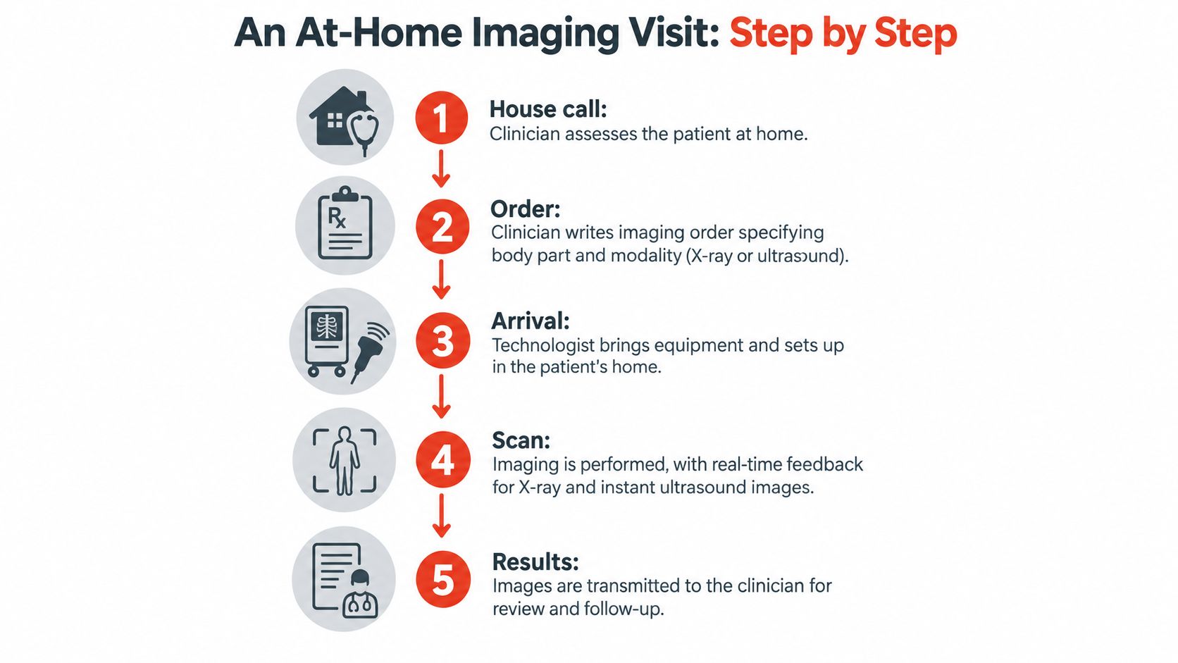
The process usually starts during a primary care house call. The clinician listens to the concern, examines the patient, and identifies a specific question. For example, the question may be whether a fall caused a fracture, whether new respiratory symptoms warrant a chest image, or whether swelling calls for an ultrasound evaluation.
The order gives the scan a purpose
The clinician writes an order naming the body part, the requested modality, and the clinical indication. That information helps the imaging team select the correct protocol and gives the radiologist the context needed to interpret the images.
Next, a scheduling coordinator confirms practical details such as the patient's address, insurance information, mobility needs, and the room where the study can take place. Routine requests may require scheduling, while urgent requests depend on clinical circumstances, availability, and local coordination. A mobile imaging service describes a similar patient workflow: obtain a physician's order, locate a provider, and have a technologist bring portable equipment to the home before sending results to the physician. Details vary among providers.
The technologist sets up around the patient
The technologist may work in a bedroom, living room, or another private space. The patient can usually remain on a bed or sit in a chair, depending on the examination and safe positioning. The study is limited to what the order requests, rather than functioning as a complete hospital radiology appointment.
Images are transmitted securely to a radiologist for review. The report then returns to the ordering clinician, who explains what the result means in relation to the symptoms and decides whether follow-up, treatment, another test, or hospital evaluation is needed. A home-health imaging provider describes bedside X-ray and ultrasound as services designed for patients who need imaging where they live, including people who are bed-bound or have limited mobility.
The scan answers one question. The clinician's job is to place that answer inside the larger story of the patient's health.
Who This Helps and Where It Falls Short
The strongest candidates for mobile imaging are people for whom transportation is difficult but who remain clinically stable enough for a home-based assessment. That can include a frail older adult after a fall, someone recovering from surgery, a person living with dementia who becomes distressed during transfers, or a patient whose oxygen, wheelchair, or caregiver needs make an outpatient visit unusually burdensome.
At-home imaging can support questions involving a possible fracture, a chest problem, fluid, selected abdominal findings, or swelling in a limb. A 2023 statistic cited in a mobile imaging market report found that portable X-ray services prevented hospital admission for approximately 81% of patients whose X-rays were normal, illustrating how a normal radiograph may help avoid unnecessary escalation when the clinician believes home management remains appropriate. That figure comes from the mobile imaging services market report, and it shouldn't be treated as a promise for an individual patient.
Where home imaging fits well
| Clinical Scenario | Best Fit at Home | Limitation to Consider |
|---|---|---|
| Fall with localized pain | Mobile X-ray may assess selected bones or joints | A normal image may not exclude every injury |
| New respiratory concern | Chest X-ray may provide useful information | Severe breathing difficulty requires urgent evaluation |
| Swollen limb | Ultrasound may assess selected vascular questions | The exam depends on the body area and operator |
| Abdominal or soft-tissue concern | Ultrasound may show selected organs, fluid, or tissue changes | Gas, body habitus, and positioning can limit the view |
| Stable patient who cannot travel | Bedside imaging avoids a difficult transfer | The study may be narrower than a hospital workup |
| Suspected stroke or major trauma | Emergency department evaluation is the appropriate setting | Mobile imaging cannot replace emergency response |
The boundaries matter
Portable systems have a smaller field of view and may need careful calibration. Ultrasound can be highly dependent on the operator and on whether sound can reach the structure of interest. A patient may still need CT, MRI, advanced laboratory testing, specialist assessment, or hospital observation.
Home imaging isn't the right response to sudden weakness, suspected stroke, severe abdominal pain, major trauma, severe breathing trouble, or rapid deterioration. In those situations, call emergency services rather than waiting for a mobile technologist.
How Life Primary Care Coordinates Mobile Imaging
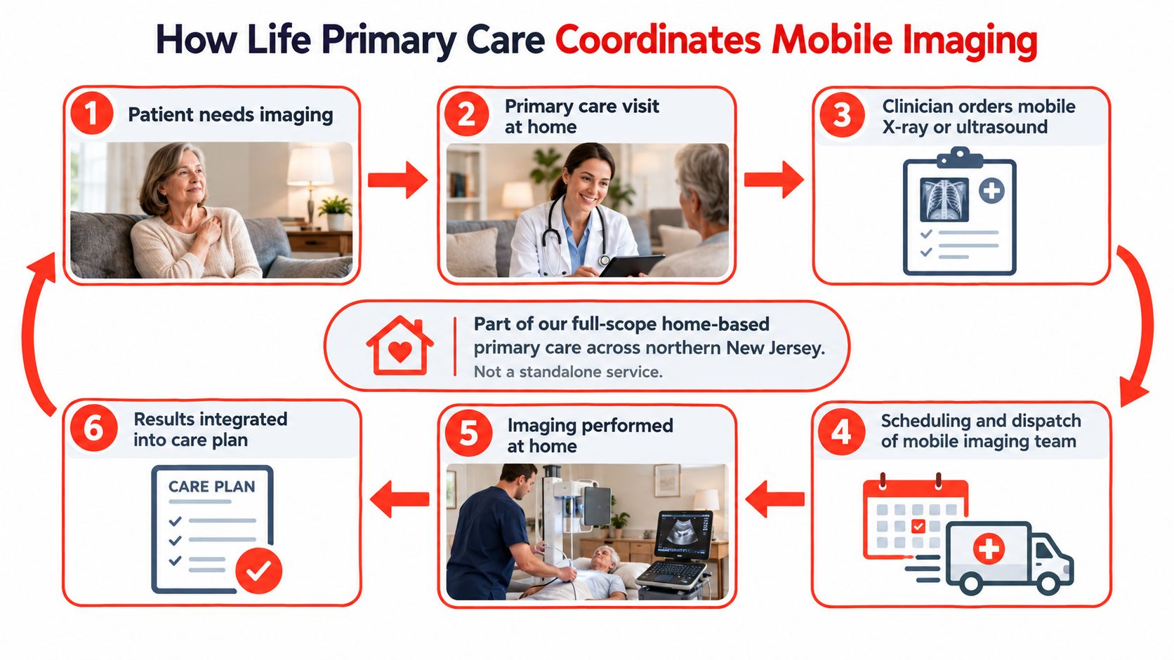
The practical difference lies in coordination. A standalone scan can produce an image, but a primary care house call begins by asking what decision the image needs to support. During a 45 to 60 minute home visit, a clinician can take a history, perform a physical examination, review medication bottles, assess the home environment, and discuss the patient's goals before ordering imaging.
If a scan is warranted, Life Primary Care places the order and coordinates authorization, scheduling, and communication with the family. A credentialed mobile imaging partner then brings the appropriate portable equipment to the residence. The radiologist's report returns to the primary care clinician, rather than leaving the family to interpret technical language alone.
Why context changes the meaning of a result
Suppose an older adult has mild shortness of breath, ankle swelling, heart failure, and several medications prescribed by different clinicians. A mobile image may be useful, but the report isn't the whole assessment. The ordering clinician must connect it with the examination, recent symptoms, medication list, advance directives, and the patient's priorities.
That integration can prevent two opposite errors. A family shouldn't assume a reassuring image makes every symptom harmless, and it shouldn't panic over an incidental finding that doesn't explain the current problem. The clinician decides whether the result changes management and communicates the next step.
Life Primary Care serves patients across northern and central New Jersey, including homes, apartments, family residences, and assisted living communities. Its in-home diagnostic coordination includes mobile X-ray and ultrasound alongside services such as labs, EKGs, urine testing, chronic disease management, urgent visits, and follow-up. Details about in-home labs and diagnostics can help families understand how imaging fits into a broader home-care plan.
Clinical context matters: A portable image is most useful when the ordering clinician knows the patient, defines the question, and follows through after the report arrives.
Technology can support this coordination, but it doesn't replace the clinician's judgment. Teams exploring healthcare AI adoption services may be interested in workflow tools, while families should focus on who will review the result, explain it, and arrange care afterward.
Practical Questions Families Ask Before Booking
Families usually want to know three things first: Will it be covered, how soon can someone come, and who will explain the result? The answers depend on the ordering clinician, the imaging provider, the patient's insurance plan, the medical indication, and local availability.
Medicare Part B can cover portable X-ray, ultrasound, and EKG services when a physician orders them, according to an independent mobile-imaging service's patient guidance. Coverage still depends on the order, medical necessity, documentation, and payer rules. Travel or call-out fees deserve a direct question before scheduling because reimbursement isn't consistent across modalities and markets. In Australia, for example, one source notes a specific Medicare item number for the mobile X-ray travel component, while mobile ultrasound travel doesn't receive the same treatment. That example shows why families shouldn't assume that an imaging fee includes every travel charge.
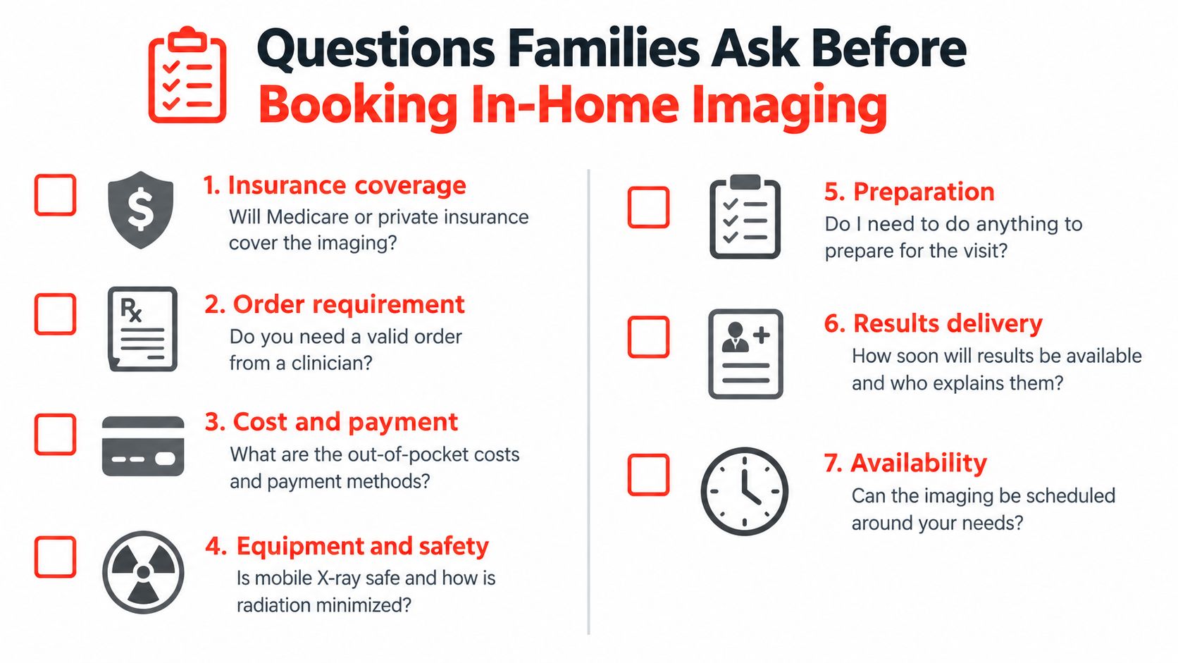
Questions to ask the coordinator
- Insurance coverage: Ask whether Medicare or private insurance covers the ordered study.
- Order requirement: Confirm that a valid clinician's order is needed and find out who sends it.
- Cost and payment: Request a clear explanation of out-of-pocket costs, travel charges, and payment methods.
- Equipment and safety: Ask how the technologist manages positioning, radiation safety, and privacy.
- Preparation: Check whether clothing, fasting, medication changes, or room preparation is necessary.
- Results delivery: Ask when the radiologist's report is expected and which clinician will explain it.
- Availability: Confirm whether the visit can be scheduled around the patient's care routine and mobility needs.
The equipment generally needs enough space to position the patient and operate safely. A coordinator can explain whether a bed, chair, outlet, or clear floor area is needed. Pets and family members may need to move temporarily during an X-ray exposure, while a caregiver can often remain nearby when the technologist says it's safe.
For families deciding whether a home visit is appropriate, this guide to whether doctors can make house calls offers useful context. Mobile imaging stops where emergency care begins. Suspected stroke, severe abdominal pain, major trauma, severe breathing difficulty, or rapid decline call for emergency services and a hospital, not a routine home scan.
Life Primary Care provides full-scope primary care at home across northern and central New Jersey, including house-call examinations, chronic disease care, urgent visits, and coordination of mobile X-ray and ultrasound when clinically appropriate. If leaving home is difficult for you or an older family member, visit Life Primary Care to learn how an at-home assessment can connect symptoms, imaging, and follow-up in one coordinated plan.
Ready for a visit at home?
Call and speak with a real person. We will confirm your insurance, find a time and come to you.
973-607-4911 Call now